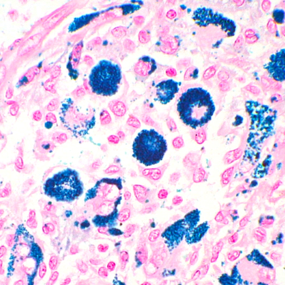
 Perls Stain satined on Giant cell tumour of tendon sheath

Perls stain method is considered to be the first classical histochemical reaction to demonstrate iron especially in tissues such as bone marrow, spleen. This procedure is particularly helpful to evaluate pathological conditions that involve hemosiderin deposits. In addition to haemorrhage, this can occur in conditions such as haemochromatosis (where excessive amounts of iron may form in organs due to iron overload) and in some liver diseases.
Perls stain method is considered to be the first classical histochemical reaction to demonstrate iron especially in tissues such as bone marrow, spleen. This procedure is particularly helpful to evaluate pathological conditions that involve hemosiderin deposits. In addition to haemorrhage, this can occur in conditions such as haemochromatosis (where excessive amounts of iron may form in organs due to iron overload) and in some liver diseases.