

Myeloproliferative neoplasms, or MPNs, are clonal disorders of hematopoietic stem and progenitor cells marked by persistent proliferation of one or more myeloid lineages. They can be difficult to pin down. Blood counts, marrow morphology, fibrosis, and molecular findings often overlap across disease categories, which means no single test tells the whole story. This is where a focused immunohistochemistry panel can help, and it's the subject of this post.
The classical BCR::ABL1 negative MPNs include polycythemia vera (PV), essential thrombocythemia (ET), and primary myelofibrosis (PMF). The broader MPN family also includes BCR::ABL1 positive chronic myeloid leukemia along with other less common entities. Because clinical findings, blood counts, marrow morphology, fibrosis, and molecular abnormalities can look similar across these categories, recognizing them accurately takes more than one line of evidence.
Modern MPN diagnosis depends on integration rather than a single test. Bone marrow morphology remains central to classification, while molecular testing for disease associated driver abnormalities and other clonal markers provides complementary evidence. Immunohistochemistry adds another layer by highlighting progenitor and blast populations along with lineage specific marrow components directly within tissue architecture.
A bone marrow biopsy holds spatial information that a blood count alone cannot capture. IHC helps the pathologist recognize the distribution and relative prominence of immature cells, megakaryocytes, granulocytic elements, and erythroid precursors. This becomes especially useful when marrow architecture is altered by hypercellularity or fibrosis, or when disease progression and blast transformation are a concern.
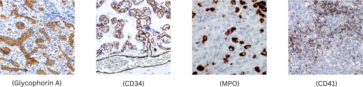
PathnSitu's focused MPN oriented marker set brings together five complementary targets: CD34 (EP88/Qbend10), CD117 (Polyclonal/EP10/PRM117), CD41 (EP178), Myeloperoxidase/MPO (EP151), and Glycophorin A (EP213). These antibodies aren't meant to define an MPN subtype on their own. Rather, they're best used as part of an integrated hematopathology panel.
Five Markers, Five Complementary Views of the Marrow
|
Marker / Clone |
What it highlights |
Potential value in marrow assessment |
|
CD34 (EP88 / Qbend10) |
Hematopoietic stem/progenitor and blast populations |
Supports assessment of immature/blast cells and possible disease progression |
|
CD117 (Polyclonal / EP10 / PRM117) |
c-KIT expressing hematopoietic progenitors and mast cells |
Complements evaluation of immature myeloid populations in context |
|
CD41 (EP178) |
Megakaryocytic lineage |
Highlights megakaryocytes and supports assessment of megakaryocytic proliferation |
|
MPO (EP151) |
Myeloid/granulocytic differentiation |
Supports recognition of myeloid lineage and granulocytic components |
|
Glycophorin A (EP213) |
Erythroid lineage and precursors |
Highlights erythroid differentiation and distribution |
CD34 is expressed during the stem/progenitor stage of hematopoietic development. In bone marrow pathology, CD34 IHC is particularly valuable for visualizing immature hematopoietic cells and estimating the distribution of blasts within the biopsy. In an established MPN, an abnormal increase in blasts may signal disease acceleration or transformation and warrants comprehensive hematopathologic assessment. PathnSitu offers both EP88 and Qbend10 CD34 clones, giving laboratories alternative validated antibody options for human FFPE tissue.
CD117, or c-KIT, is a receptor tyrosine kinase expressed across several hematopoietic cell populations, including progenitors and mast cells. In marrow IHC, CD117 can complement CD34 when evaluating immature myeloid elements, though its broader expression means interpretation must stay morphologic and context dependent. PathnSitu provides multiple CD117 options, Polyclonal, EP10, and PRM117, so laboratories can select an antibody configuration suited to their validated workflow.
Megakaryocytic morphology sits at the center of the distinction among several MPN categories. CD41 is expressed on platelets and megakaryocytes and is widely used as a marker of megakaryocytic differentiation. In marrow sections, CD41 makes the megakaryocytic compartment easier to appreciate, helping the observer evaluate its distribution and proliferation alongside H&E morphology and fibrosis assessment.
Myeloperoxidase is a major component of the azurophilic granules of neutrophilic granulocytes and is a well established marker of myeloid differentiation. MPO IHC helps visualize the granulocytic/myeloid compartment and provides useful lineage information when immature myeloid cells are under evaluation. PathnSitu's EP151 clone is listed for human FFPE tissue and shows cytoplasmic localization.
Glycophorin A (CD235a) is an erythroid associated membrane protein expressed on erythroid cells and their precursors. Its inclusion allows the erythroid compartment to be evaluated alongside granulocytic and megakaryocytic lineages, which is particularly informative when assessing trilineage proliferation or altered erythroid distribution within a marrow biopsy.
CD34/CD117 → immaturity | CD41 → megakaryocytic lineage | MPO → myeloid lineage | Glycophorin A → erythroid lineage
Current classification frameworks emphasize integrating bone marrow morphology with blood findings and molecular genetics. In PV, ET, and PMF, testing for JAK2, CALR, and MPL mutations can provide evidence of clonality and support subtype designation, but molecular results do not replace morphologic evaluation. IHC should be viewed the same way. It enhances recognition of specific cellular compartments and can clarify difficult morphology, but it does not independently establish the complete MPN diagnosis.
A practical workflow combines complete blood counts and peripheral smear review, bone marrow morphology and fibrosis grading, a focused IHC panel, and appropriate molecular/cytogenetic testing. When these data are read together, the pathologist gains a more complete view of lineage proliferation, marrow architecture, clonality, and possible progression.
A high quality diagnostic result depends not only on selecting the right antibody, but also on the consistency of the entire IHC process. PathnSitu's broader tissue diagnostics approach connects antigen retrieval, primary antibodies, automated staining, and digital visualization into one laboratory workflow.
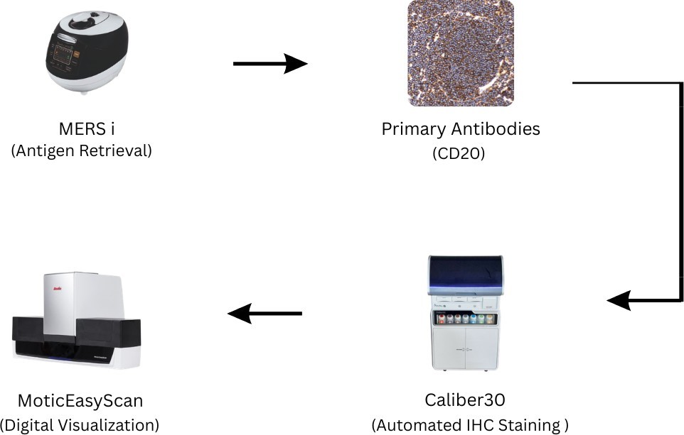
Consistent epitope retrieval is an important pre-analytical step for FFPE IHC. A validated retrieval workflow helps prepare tissue sections for reproducible antibody staining and reduces avoidable variability before the primary antibody is applied.
The combination of CD34, CD117, CD41, MPO, and Glycophorin A provides a compact lineage oriented panel for evaluating immature, megakaryocytic, myeloid, and erythroid components of the marrow. Marker selection should always be tailored to the morphology and clinical question.
Standardized automated staining helps laboratories manage multi-marker IHC workflows with reduced hands-on variability. PathnSitu's automation portfolio is built to support consistent staining and laboratory efficiency as part of its broader tissue diagnostics ecosystem.
Digital microscopy and slide scanning support visualization, documentation, review, and consultation of stained marrow sections. The final interpretation remains an integrated professional judgment based on morphology, IHC staining patterns, clinical information, and appropriate ancillary studies.
PathnSitu's differentiator is best described as an integrated, laboratory focused approach rather than a claim that one antibody is inherently superior to every competing product. Its portfolio combines multiple clone options for selected targets, antibodies available for FFPE workflows, and a broader emphasis on streamlining staining and turnaround time. For this MPN focused panel, laboratories can access two CD34 clones (EP88 and Qbend10), three listed CD117 options (Polyclonal, EP10, and PRM117), together with lineage oriented CD41 EP178, MPO EP151, and Glycophorin A EP213. This breadth gives laboratories flexibility to build and validate a panel around their own tissue, platform, and protocol requirements. PathnSitu also positions its tissue diagnostics offering around quality, technical support, workflow efficiency, and affordability, practical considerations that matter when a laboratory is seeking a sustainable diagnostic solution.
MPNs show why modern hematopathology increasingly depends on integration. The same marrow may need assessment of megakaryocytic architecture, erythroid and granulocytic proliferation, fibrosis, immature cell burden, and molecular evidence of clonality. No single stain answers all of these questions.
PathnSitu's CD34, CD117, CD41, MPO, and Glycophorin A antibodies provide complementary visual information across key marrow compartments. Used with careful morphology, clinical data, and appropriate molecular testing, they can strengthen the pathologist's ability to recognize abnormal patterns and direct the next diagnostic step.
PathnSitu: Better visualization of lineage. Better integration of evidence. A clearer path from marrow morphology to diagnostic insight.
Reference