

High-Quality Staining Is Critical for Accurate Diagnosis
When "Just Appendicitis" Turns Out to Be Cancer
Appendix cancer is rare - and it hides well. Under a microscope, it can look almost identical to colon cancer, ovarian cancer, or a simple inflamed appendix. Get the staining wrong, and a patient risks the wrong surgery or a dangerous delay in treatment.
How the Right Stains and Antibodies Confirm an Appendix Cancer Diagnosis
Because appendix tumors are so diverse, pathologists need more than one stain to be sure. PathnSitu Biotechnologies supports this diagnostic process with three tools working together.
Consistency Matters: From Antigen Retrieval to Automated IHC
Accurate antibodies are only part of the equation. Reliable IHC results also depend on consistent tissue preparation, antigen retrieval, reagent application, and incubation. For laboratories handling rare and diagnostically challenging appendix tumors, controlling these variables can help reduce staining variability and support confident interpretation.
MERS: Consistent Antigen Retrieval for Manual IHC Workflows
Before an antibody can bind to its target, tissue antigens may need to be exposed through controlled heat- and pressure-based antigen retrieval. When performed manually, differences in temperature, pressure, timing, or protocol execution can affect staining quality.
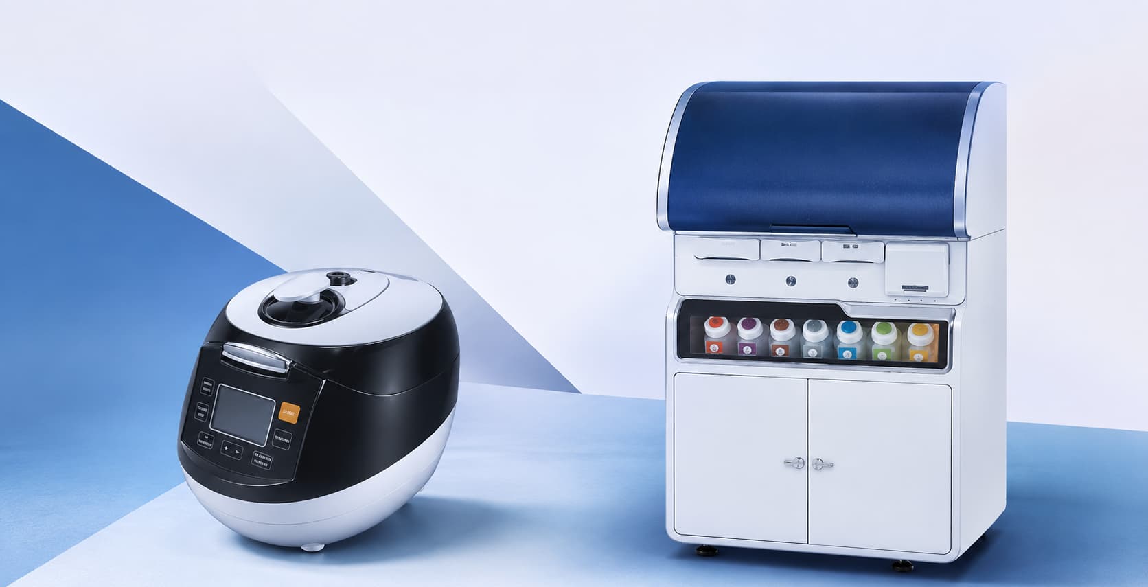
MERS helps standardize this critical preparation step with a pressure-based walkway system and digitally controlled protocols. Features such as one-touch programs, temperature display and control, on-demand pressure cycles, and multiple protocols in a single run give laboratories greater control over antigen retrieval conditions. With a capacity of up to 72 slides per run, MERS can also support higher-throughput manual IHC workflows.
For appendix cancer markers such as SATB2, CK20, CDX2, and Chromogranin A, consistent antigen retrieval can help produce clear, reproducible staining that pathologists can interpret with greater confidence.
Caliber 30: Automating the IHC Staining Process
Once tissue preparation is standardized, the next challenge is delivering the IHC staining process consistently from slide to slide. This is where Caliber 30, a fully automated IHC staining system, fits into the workflow.
Caliber 30 automates the IHC process from baking through counterstaining, reducing the hands-on variability associated with manual staining. Its on-board antigen retrieval uses proven technology, while features such as full-slide coverage with 120 µL and on-board DAB mixing help maintain controlled and reproducible staining across a batch.
For a diagnostic panel involving markers such as SATB2, CK20, CDX2, and Chromogranin A, automation helps ensure that reagent application, processing steps, and staining conditions are consistently controlled.
In the end, this is what the automated workflow is really solving for: giving pathologists consistent, repeatable results they can trust.