

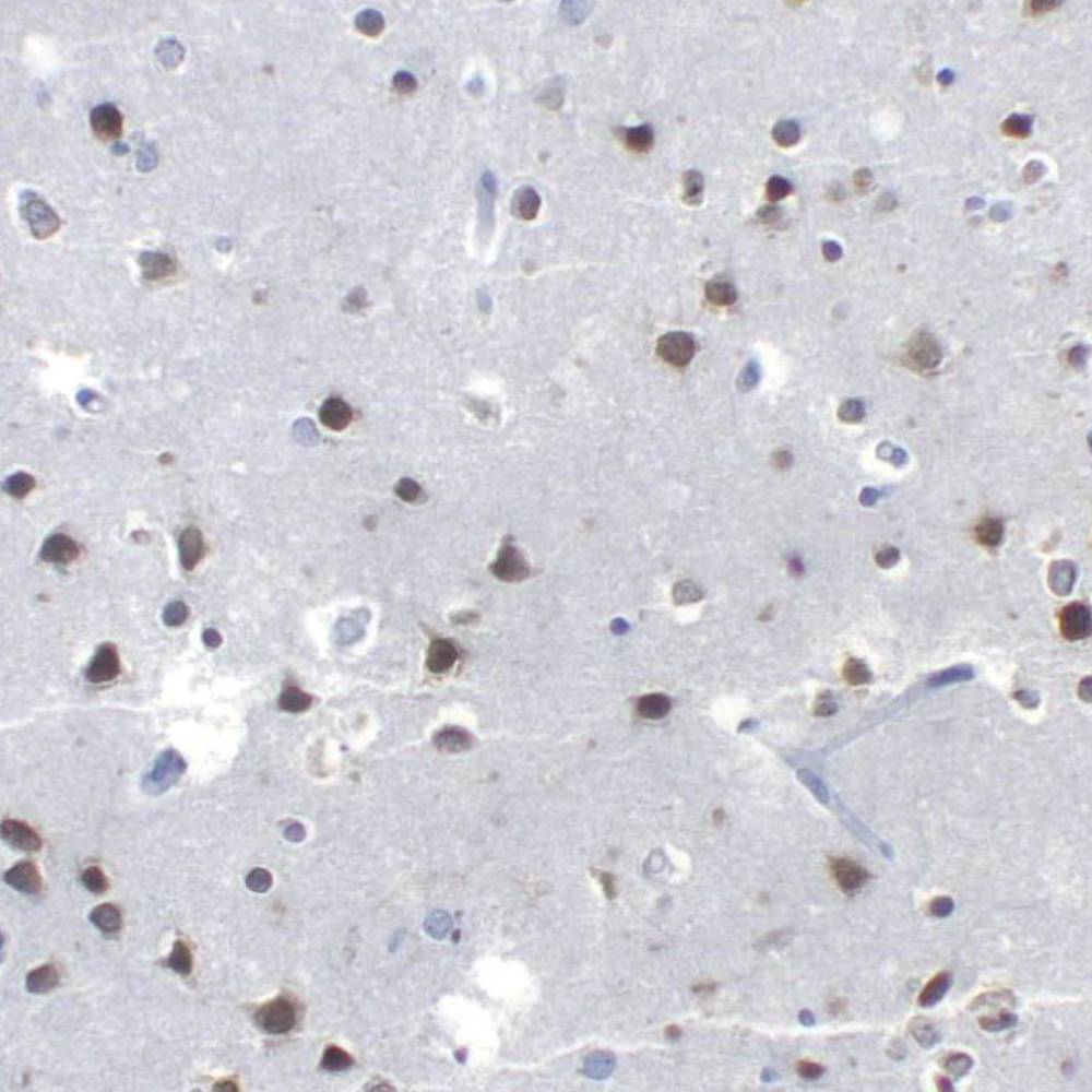
 Brain stained with Anti-NeuN
NeuN antibody specifically recognizes the DNA-binding, neuron-specific protein NeuN, which is present in most CNS and PNS neuronal cell types of all vertebrates tested. NeuN protein distributions are apparently restricted to neuronal nuclei, perikarya and some proximal neuronal processes in both fetal and adult brain although, some neurons fail to be recognized by NeuN at all ages: INL retinal cells, Cajal-Retzius cells, Purkinje cells, inferior olivary and dentate nucleus neurons, and sympathetic ganglion cells are examples. Immunohistochemically detectable NeuN protein first appears at developmental timepoints that correspond with the withdrawal of the neuron from the cell cycle and/or with the initiation of terminal differentiation of the neuro. Immunoreactivity appears around E9.5 in the mouse neural tube and is extensive throughout the developing nervous system by E12.5. Strong nuclear staining suggests a nuclear regulatory protein function; however, no evidence currently exists as to whether the NeuN protein antigen has a function in the distal cytoplasm or whether it is merely synthesized there before being transported back into the nucleus. No difference between protein isolated from purified nuclei and whole brain extract on immunoblots has been found.
NeuN antibody specifically recognizes the DNA-binding, neuron-specific protein NeuN, which is present in most CNS and PNS neuronal cell types of all vertebrates tested. NeuN protein distributions are apparently restricted to neuronal nuclei, perikarya and some proximal neuronal processes in both fetal and adult brain although, some neurons fail to be recognized by NeuN at all ages: INL retinal cells, Cajal-Retzius cells, Purkinje cells, inferior olivary and dentate nucleus neurons, and sympathetic ganglion cells are examples. Immunohistochemically detectable NeuN protein first appears at developmental timepoints that correspond with the withdrawal of the neuron from the cell cycle and/or with the initiation of terminal differentiation of the neuro. Immunoreactivity appears around E9.5 in the mouse neural tube and is extensive throughout the developing nervous system by E12.5. Strong nuclear staining suggests a nuclear regulatory protein function; however, no evidence currently exists as to whether the NeuN protein antigen has a function in the distal cytoplasm or whether it is merely synthesized there before being transported back into the nucleus. No difference between protein isolated from purified nuclei and whole brain extract on immunoblots has been found.


